Routine imaging is worthwhile in follow-up of high-risk melanoma patients

Background There is no international consensus on follow-up imaging in cutaneous melanoma. The aim of our study was to examine whether melanoma recurrences were detected by routine follow-up imaging or by symptom-based imaging.
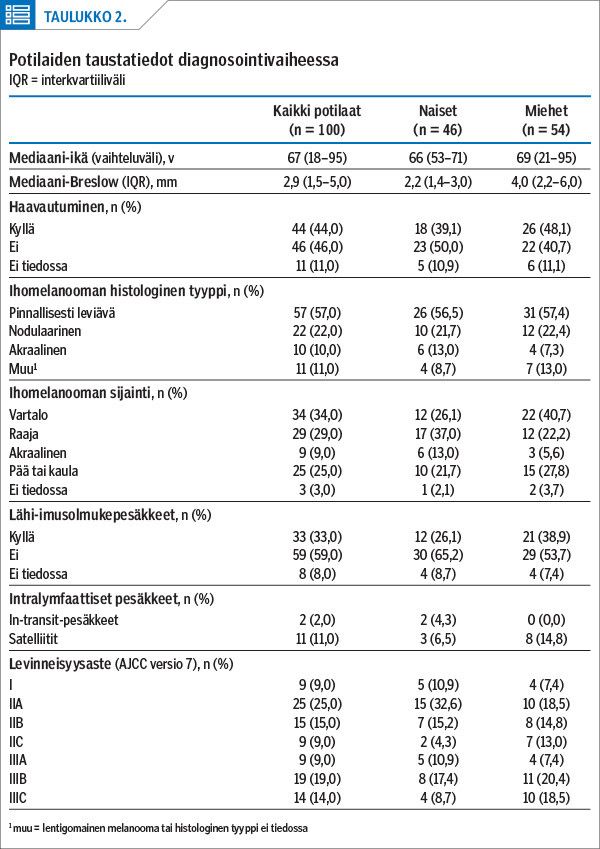
Methods We analysed the methods of recurrence detection for 100 stage I–III melanoma patients referred to the Department of Oncology at Tampere University Hospital during 2012–2014.
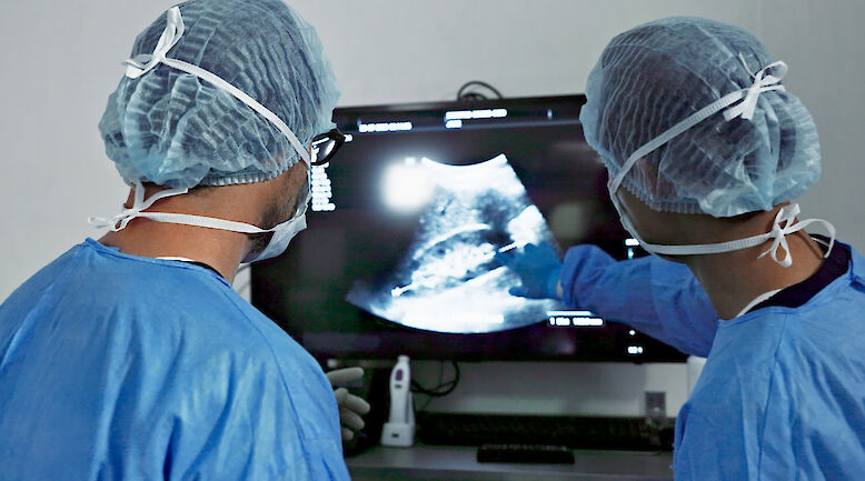
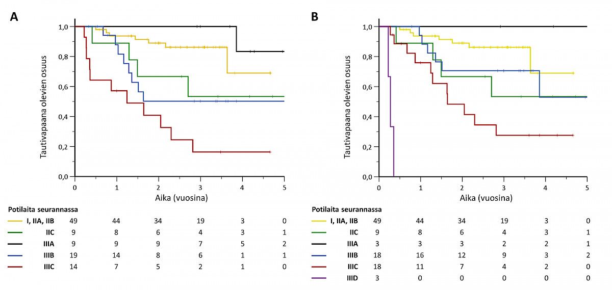
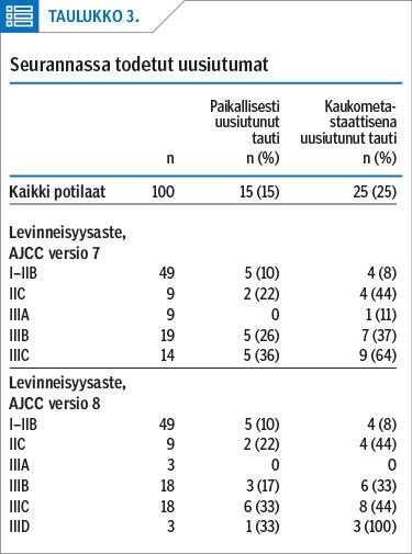
Results One third of high-risk melanoma patients had a relapse during the first three years of follow-up. Half (50%) of all the melanoma recurrences were detected by routine imaging and 35% by symptom-based imaging. In the case of systemic recurrences 60% were identified by routine imaging and only 28% by symptoms. Locoregional relapses were detected almost equally by routine and symptom-based imaging (38% vs. 42%). In terms of imaging modalities, systemic relapses were most commonly detected by CT scans (72%) and locoregional recurrences by lymph node ultrasound examinations (52%).
Conclusions Due to the considerable number of recurrences follow-up of stage IIB-III melanoma patients in specialized medical care is justified. New treatment strategies have improved survival in metastatic melanoma. Routine CT imaging and regional US examinations are recommended for high-risk melanoma patients in order to detect recurrences earlier.